Have you been searching for what is Wrinkles the Clown’s phone number after hearing stories about this creepy character? Wrinkles the Clown became an internet sensation and local legend in Florida, with parents reportedly hiring him to scare their misbehaving children. While a phone number was once circulated online, the story behind Wrinkles is much more complex and interesting than you might think. This guide will explore the truth about Wrinkles the Clown, the famous phone number, what really happened when people called it, and the documentary that revealed the surprising reality behind this viral phenomenon.
Who Is Wrinkles the Clown?
Wrinkles the Clown first appeared in 2014 in Southwest Florida, particularly around Naples and Fort Myers. The character was a person dressed in a creepy clown costume, complete with a wrinkled mask, who would show up to “scare” children whose parents hired him.
The concept was simple but unsettling: if your child was misbehaving, you could supposedly call Wrinkles, and he would show up at your house to frighten your child into better behavior. He would stand outside windows, emerge from under beds, or appear in unexpected places.
What made Wrinkles different from typical scary stories was that he seemed real. There were videos, a phone number you could actually call, and local news reports. Parents in Florida claimed they’d really hired him. The combination of creepy clown imagery and the ability to actually contact this character made Wrinkles go viral.
The character played on common childhood fears – clowns, things under the bed, and faces at the window. But unlike Slenderman or other internet horror creations, Wrinkles appeared to exist in the real world, which made him even more terrifying to children and fascinating to adults.
The Famous Phone Number
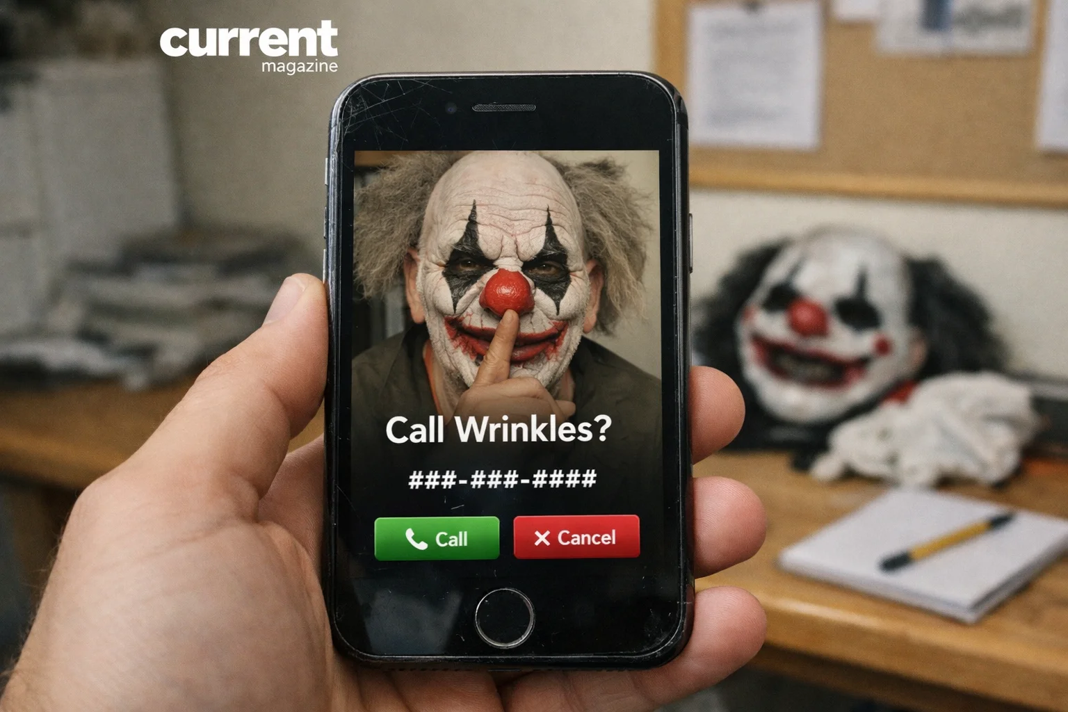
The phone number associated with Wrinkles the Clown was (941) 365-4935. This number circulated widely online, in news reports, and across social media platforms.
When the number was active, people who called it would reach a voicemail message. The recording featured a creepy, distorted voice that would say something along the lines of: “Hello, you’ve reached Wrinkles. Leave a message with your child’s name and address, and I’ll make a visit.”
Thousands of people called this number out of curiosity, to prank friends, or allegedly to actually hire Wrinkles for a scare. The voicemail box would fill up with messages ranging from:
- Curious people asking if this was real
- Terrified children begging Wrinkles not to come to their house
- Parents pretending to hire him to scare their kids
- Pranksters leaving joke messages
- True crime enthusiasts investigating the phenomenon
Important note: This phone number is no longer active and has not been for several years. Calling it today will either not connect or reach a different person/business entirely. Do not call random numbers expecting to reach Wrinkles – the phenomenon has been over since around 2016-2017.
The Videos That Made Wrinkles Famous
What really propelled Wrinkles to viral status were the videos posted online. These showed:
Wrinkles appearing outside children’s windows at night, with kids screaming and crying when they saw the clown face pressed against the glass.
Wrinkles emerging from under beds, where he’d supposedly been hiding, to terrify children who thought monsters under the bed were just stories.
Wrinkles standing in yards or driveways, silently staring at houses, creating an atmosphere of dread.
Parents filming their children’s reactions, documenting the moment their kids saw Wrinkles, which raised serious questions about the ethics of deliberately terrifying children.
These videos accumulated millions of views on YouTube, Facebook, and other platforms. They sparked heated debates: Was this real? Was it child abuse? Was it effective parenting or psychological harm? Was the whole thing a hoax?
The grainy, amateur quality of many videos made them seem authentic, like real security camera footage or genuine phone recordings, which added to the mystery and fear factor.
The Documentary That Revealed the Truth
In 2019, a documentary titled “Wrinkles the Clown” premiered, directed by Michael Beach Nichols. This film investigated the phenomenon and ultimately revealed surprising truths about the person behind the mask.
What the documentary uncovered:
The person behind Wrinkles was revealed to be a 65-year-old man named Jack Ryals (though some sources spell it differently). He lived in Southwest Florida and came up with the Wrinkles concept as what he described as a “performance art project.”
The big reveal: Most of the scariest videos weren’t real. Ryals admitted that he staged many of the viral videos using actors – both adults playing parents and children who were acting. The videos that showed the most dramatic reactions, the ones that went most viral, were largely fabricated for shock value.
However, some aspects were real. The phone number was genuine, and Ryals did receive thousands of actual calls. He did make some actual “appearances,” though the documentary suggests these were more photoshoots and public sightings rather than true home visits to scare children.
Why did he do it? According to the documentary, Ryals was interested in exploring internet culture, viral phenomena, and how easily people believe what they see online. He was also making commentary on parenting, fear, and the dark side of social media.
The documentary showed Ryals as a somewhat eccentric but ultimately harmless older man who stumbled into creating a viral sensation that grew far beyond what he originally intended.
Was Wrinkles Real or a Hoax?
The answer is: both and neither, depending on what you mean by “real.”
What was real:
- A person named Jack Ryals did dress as Wrinkles the Clown
- The phone number (941) 365-4935 was real and functional
- People really did call and leave messages
- Some public appearances and sightings genuinely occurred
- Local news really did cover the story
- The character became a genuine cultural phenomenon in Southwest Florida
What wasn’t real:
- Most of the viral videos showing children being terrified were staged with actors
- The idea that numerous parents regularly hired Wrinkles to scare their children was exaggerated
- Many of the scariest stories were fabrications or urban legends that grew around the real core
- The voicemail messages from “terrified children” included many pranks and people playing along
So Wrinkles existed as a real performance art character, but the narrative around him – the scary clown-for-hire business – was largely constructed for viral impact rather than being a genuine service.
The Ethics and Controversy
Wrinkles the Clown sparked significant ethical debates, particularly around child psychology and parenting:
Arguments that it was harmful:
- Deliberately terrifying children can cause lasting psychological trauma
- Using fear as a parenting tool teaches children that the world is dangerous and that parents can’t be trusted
- Young children can’t distinguish between real threats and pretend ones
- It could trigger anxiety, nightmares, and phobias
- The videos potentially exploited children’s genuine fear for entertainment and views
Arguments defending it:
- Most dramatic videos were staged, so no real children were harmed
- Some parents argued controlled fear (like haunted houses) is normal childhood experience
- It was performance art commentary on modern parenting and internet culture
- The phenomenon was mostly urban legend rather than widespread practice
What child psychologists said: Mental health experts who weighed in generally agreed that deliberately terrifying young children, even as discipline, is psychologically harmful and ineffective. Fear-based parenting can damage parent-child trust and doesn’t teach children why behaviors are wrong or how to make better choices.
Whether real or mostly staged, Wrinkles the Clown highlighted concerning trends in viral content – the willingness to frighten children for views and the blurry line between entertainment and harm online.
Why Wrinkles Went Viral
What made this particular creepy clown capture the internet’s imagination?
Perfect timing: Wrinkles emerged during peak “creepy clown” culture. In 2014-2016, there was a nationwide “clown scare” with reports of threatening clowns appearing in various states. This created a cultural moment where people were primed to fear and be fascinated by scary clowns.
The accessibility factor: Unlike other horror legends, you could supposedly contact Wrinkles directly. That phone number made it feel real and interactive.
Shocking parenting angle: The idea that parents would hire someone to terrorize their own children was so outrageous it demanded attention and sparked heated debates.
Local news legitimacy: When local Florida news stations covered Wrinkles, it added credibility. If it was on the news, it seemed more real than just another internet story.
Viral video format: The short, sharable videos were perfect for social media. They were just long enough to be scary but short enough to go viral.
Mystery and ambiguity: For years, no one knew who was behind the mask or whether it was real, which kept people talking and investigating.
What Happened to Wrinkles?
After the 2019 documentary revealed the truth behind Wrinkles, the phenomenon largely ended.
The phone number was disconnected and is no longer in service. You cannot call and reach Wrinkles anymore.
Jack Ryals essentially retired the character once the documentary exposed the reality behind it. The mystery was gone, and with it, much of the fear and fascination.
Copycat Wrinkles appeared in various places, with other people trying to recreate the phenomenon, but none achieved the same viral status as the original.
The character remains in internet history as a notable example of viral culture, urban legends, and the intersection of horror and social media.
Today, Wrinkles the Clown is more of a cultural footnote – a weird internet phenomenon from the mid-2010s that briefly terrified children and fascinated adults before being revealed as largely performance art.
Similar Phenomena and Urban Legends
Wrinkles wasn’t the first or last scary character to capture public imagination:
Slenderman: A tall, faceless figure created on internet forums that became so widely believed that it led to real-world violence when two girls attacked a classmate, claiming Slenderman made them do it.
The Bye Bye Man: An urban legend turned horror movie about a supernatural entity you summon by saying his name.
Momo Challenge: A supposed social media challenge encouraging children to self-harm, which turned out to be largely a moral panic with little evidence of actual danger.
Killer Clown Scare of 2016: Reports across the US of creepy clowns lurking in woods and neighborhoods, which were mix of pranks, hoaxes, and mass hysteria.
What they all share: These phenomena blend online culture with real-world fears, creating modern folklore that spreads through social media rather than campfire stories.
How to Talk to Kids About Scary Internet Content
If your child has heard about Wrinkles or similar scary internet legends, here’s how to address their fears:
Acknowledge their feelings: Don’t dismiss their fear as silly. To them, it feels very real.
Explain what’s real and what’s not: Use age-appropriate language to explain that Wrinkles was mostly a made-up character, like a movie or story, not a real danger.
Discuss internet reliability: Teach kids that not everything online is true, and people sometimes create fake scary content for attention or entertainment.
Reassure them about safety: Let them know your home is safe, you wouldn’t let anyone harm them, and scary internet stories aren’t real threats.
Monitor their content: Be aware of what your children are watching online. Horror content marketed to kids (like scary challenges or legends) can be genuinely distressing.
Don’t use fear as discipline: The Wrinkles phenomenon should serve as a reminder that fear-based parenting can backfire and cause real anxiety.
Lessons from the Wrinkles Phenomenon
What can we learn from Wrinkles the Clown’s rise and fall?
Not everything online is real: Even videos, phone numbers, and news coverage don’t guarantee authenticity. Critical thinking is essential.
Viral content can have real impact: Even though Wrinkles was mostly performance art, real children were frightened by the stories and videos.
Fear spreads faster than facts: The scary story went viral instantly; the truth took years and a documentary to emerge.
Modern folklore evolves quickly: Wrinkles went from unknown to viral legend to exposed hoax in just a few years – much faster than traditional urban legends.
The ethics of viral content matter: Creating content that frightens children raises serious questions, even if the ultimate purpose is art or commentary.
Mystery drives engagement: Much of Wrinkles’s viral success came from the unknown. Once the documentary revealed the truth, interest faded quickly.
10 Frequently Asked Questions About Wrinkles the Clown
- Is Wrinkles the Clown’s phone number still active?
No, the phone number (941) 365-4935 that was associated with Wrinkles the Clown is no longer in service and has been disconnected for several years. Do not attempt to call this number, as it may now belong to a different person or business entirely. - Was Wrinkles the Clown real or completely fake?
Wrinkles was real in the sense that a person named Jack Ryals created and performed as the character, and the phone number existed. However, most of the viral videos showing terrified children were staged with actors, and the “business” of hiring him to scare kids was largely exaggerated for viral effect. - Did parents really hire Wrinkles to scare their children?
While some parents may have contacted the number as a prank or threat to children, the widespread narrative of parents regularly hiring Wrinkles was mostly fabricated. The documentary revealed that the most viral incidents were staged, and actual “scare visits” were rare or non-existent. - Who was the person behind Wrinkles the Clown?
The 2019 documentary “Wrinkles the Clown” revealed the person behind the mask was Jack Ryals, a 65-year-old man from Southwest Florida who created Wrinkles as a performance art project and social commentary on internet culture and parenting. - Where did Wrinkles the Clown operate?
Wrinkles was primarily associated with Southwest Florida, particularly the Naples and Fort Myers areas. While the legend spread nationally through social media, the character’s activities and sightings were concentrated in Florida’s Gulf Coast region. - Is there a Wrinkles the Clown movie or documentary?
Yes, there is a 2019 documentary titled “Wrinkles the Clown” directed by Michael Beach Nichols. This film investigates the phenomenon and ultimately reveals the truth about who created the character and why. It’s available on various streaming platforms and provides the most comprehensive look at the Wrinkles story. - Could Wrinkles the Clown hurt children psychologically?
Child psychologists generally agree that deliberately terrorizing children, even as discipline or entertainment, can cause genuine psychological harm including anxiety, nightmares, trust issues, and phobias. While most Wrinkles videos were staged, the concept itself raised serious ethical concerns about fear-based parenting. - When did Wrinkles the Clown first appear?
Wrinkles the Clown first emerged in 2014 in Southwest Florida. The phenomenon peaked between 2014-2016 during the nationwide “creepy clown” scare, and largely ended after the 2019 documentary revealed the truth behind the character. - Can I still hire Wrinkles the Clown?
No, you cannot hire Wrinkles the Clown. The phone number is disconnected, and Jack Ryals has essentially retired the character since the documentary exposed the reality behind it. Any current “Wrinkles” would be copycats, not the original. - Why did people believe Wrinkles the Clown was real?
Wrinkles seemed real because of multiple factors: an actual working phone number, videos that appeared authentic, local news coverage, the timing during the 2016 clown scare phenomenon, and the deliberately ambiguous nature of the character. The creator intentionally blurred the lines between reality and fiction, making it convincing until the documentary revealed the truth.